General Dentistry
Intraoral scanner accuracy: key factors affecting digital precision
Intraoral scanner accuracy is not a single value. It has two independent components: trueness and precision. Trueness measures how closely the digital model matches the actual intraoral geometry.
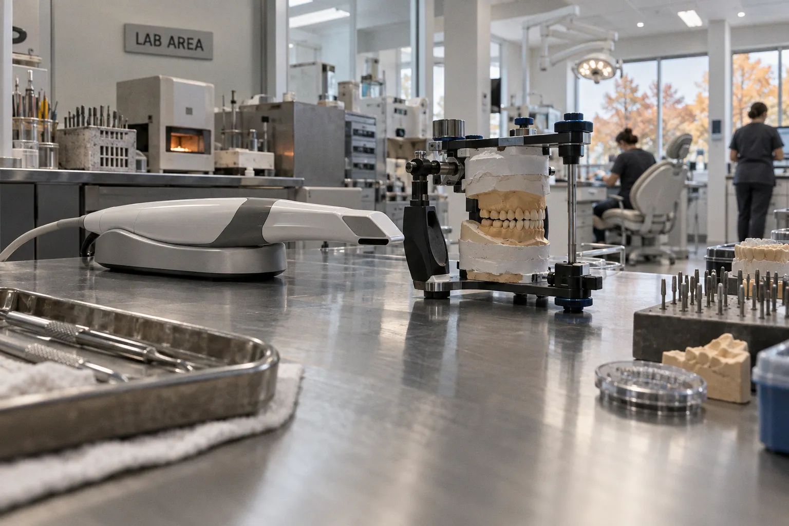
Precision measures how consistently the scanner reproduces the same geometry across repeated scans.
For high-performing systems, a practical benchmark is ≤20 μm for single-tooth scans and ≤30 μm for full-arch scans. These values are not universal clinical guarantees. They depend on scanner hardware, image-stitching software, scanning strategy, operator technique, ambient lighting, moisture control, arch morphology, and the optical behavior of restorative materials.
Digital impressions are therefore not equivalent to conventional impressions with a camera attached. They are reconstructed datasets. Every frame must be acquired, interpreted, aligned, and merged. Errors can enter at each stage.
Trueness measures deviation from anatomy. Precision measures repeatability. A scan can be precise without being true.
Defining the metrics: trueness and precision in digital dentistry
The terms are often used interchangeably. That is incorrect.
Trueness
Trueness describes systematic deviation from a reference geometry. The reference may be a master model, a calibrated physical object, or a high-accuracy laboratory dataset. The scanner output is compared with this reference through three-dimensional surface analysis.
A scan with poor trueness contains geometric bias. The entire digital model may be wider, narrower, longer, or displaced relative to the actual anatomy. On a prepared tooth, the error can affect:
- finish-line position;
- axial wall geometry;
- occlusal reduction;
- interproximal clearance;
- emergence profile;
- contact location;
- spatial relationship between the preparation and adjacent structures.
Trueness is particularly relevant when the digital model is used for CAD/CAM crown design, implant planning, guided surgery, or full-arch prostheses. A systematic geometric error can be transferred directly into the restoration or surgical guide.
Precision
Precision measures reproducibility. The same region is scanned repeatedly under equivalent conditions. The datasets are then compared with one another.
A scanner with high precision generates similar results during repeated acquisitions. This does not confirm that the geometry is correct. The system may reproduce the same distortion each time. In statistical terms, precision reflects low dispersion. Trueness reflects low deviation from the reference.
The distinction has direct procedural consequences:
| Metric | Measurement target | Typical failure pattern | Clinical implication |
|---|---|---|---|
| Trueness | Agreement with actual anatomy | Systematic dimensional deviation | Restoration or guide may be designed from incorrect geometry |
| Precision | Reproducibility between repeated scans | Variable local or cumulative distortion | Results are unstable across acquisitions |
| Combined accuracy | Trueness and precision together | Low agreement and high variability | Digital impression cannot be reliably transferred to CAD/CAM |
| Local accuracy | Specific tooth or segment | Margin, contact, or occlusal distortion | Single-unit restoration may be affected |
| Global accuracy | Entire arch or scan field | Cumulative stitching error | Full-arch prosthesis or implant relationship may be compromised |
A single reported accuracy value is therefore insufficient. Any technical assessment should identify the metric, scan length, reference method, arch condition, scanner model, software version, and scanning protocol.
Why scan length changes the error profile
Single-tooth scanning and full-arch scanning are different technical problems.
A single-tooth scan contains a limited number of image transitions. The software has fewer opportunities to accumulate registration errors. The scan field also contains local geometric features that help the algorithm maintain alignment.
A full-arch scan requires sequential registration across a longer surface. Each new image is aligned to preceding data. Small local errors can accumulate over the scan path. The final posterior segment may show a larger global deviation than the initial segment, even when every individual frame appears acceptable.
This is one reason the ≤20 μm single-tooth benchmark cannot be transferred directly to a full-arch workflow. The relevant benchmark for a full arch is less stringent in absolute terms because the reconstruction problem is more complex. The 30 μm benchmark remains a reference for top-performing systems, not a universal result under every clinical condition.
The hardware and software equation
The optical unit and reconstruction algorithm operate as a single measurement system. Hardware specifications alone do not establish clinical accuracy.
Optical hardware
Intraoral scanners acquire surface data through optical capture. The system must detect surface geometry, recognize overlapping regions, and maintain registration as the scanner moves.
Hardware variables include:
- optical resolution;
- sensor configuration;
- depth of field;
- field of view;
- scanner-tip dimensions;
- image acquisition rate;
- illumination design;
- focal behavior;
- thermal stability;
- calibration stability.
A smaller scanner tip can improve access to posterior and interproximal regions. It can also reduce the visible field captured in each frame. The software then receives less overlapping anatomy per image. This may increase the demand on scanning-path control.
A larger field of view can expose more anatomy per frame. It may simplify registration across broad surfaces. However, access limitations and soft-tissue interference can reduce the effective value of that wider field.
Scanning speed has the same non-linear relationship with accuracy. Faster acquisition can improve workflow throughput. It does not automatically improve resolution or precision. Rapid scanning may reduce the number of useful frames, shorten the time available for stable image capture, and increase motion-related registration errors.
The relevant variable is not the maximum frame rate. It is the quality of the reconstructed surface under the actual intraoral conditions.
Image-stitching algorithms
The scanner does not capture one continuous three-dimensional impression. It captures sequential datasets and combines them through image registration.
The software searches for common geometric features between adjacent frames. It then estimates the position of each new frame relative to the existing model. This process is vulnerable when the scanned area lacks stable reference features.
Registration becomes less reliable on:
- smooth, featureless surfaces;
- large uniform restorations;
- reflective alloys;
- translucent ceramics;
- wet enamel;
- moving soft tissue;
- blood-contaminated fields;
- areas with limited overlap;
- repeated or ambiguous surface patterns.
The algorithm may still produce a complete model. Completeness does not prove geometric fidelity. A visually uninterrupted surface can contain local warping or global drift.
Software updates can also change scan behavior without changing the optical hardware. Altered reconstruction algorithms may affect void filling, noise suppression, automatic trimming, occlusal registration, or the treatment of ambiguous surfaces. Scanner performance should therefore be evaluated as a hardware-software configuration, not as a permanent property of the device name.
Artificial intelligence and automated reconstruction
AI-assisted reconstruction can improve the handling of incomplete or noisy data. It can identify anatomy, suppress irrelevant tissue, classify surfaces, and estimate missing geometry. These functions may reduce visible artifacts and improve workflow speed.
They do not remove the underlying measurement problem.
An algorithm can infer a plausible surface where the scanner failed to acquire reliable data. Inferred geometry is not equivalent to directly measured geometry. The distinction matters at finish lines, implant interfaces, deep subgingival margins, and areas with weak optical contrast.
AI can also alter the operator’s perception of error. A smooth rendered model may appear clinically complete while containing reconstructed regions with lower evidentiary support. Quality control must therefore include raw acquisition behavior, not only the final polished visualization.
The unresolved comparison is the relative contribution of operator experience and AI-assisted reconstruction in the newest scanner models. Current evidence does not justify assigning a universal weight to either variable.
Clinical variables that alter digital impression quality
The optical environment inside the mouth is unstable. Moisture, lighting, anatomy, and material composition can alter the quality of captured data.
Moisture and contamination
Saliva produces a variable optical surface. It can create glare, obscure preparation margins, and change local surface texture between frames. A thin film may appear insignificant on the screen while affecting edge detection and registration.
Blood and crevicular fluid create a larger problem at subgingival margins. They can mask the finish line and create an incomplete transition between tooth structure and soft tissue. The scanner may capture a surface, but the captured surface may not represent the intended margin.
Moisture control is therefore part of the measurement protocol. It is not only a comfort or visibility measure. Retraction, air drying, suction, isolation, and tissue management determine whether the optical system can identify the target geometry.
Subgingival preparation margins remain a high-risk region because the scanner must acquire a narrow geometric boundary near mobile or reflective tissue. Margin depth, tissue displacement, fluid control, and scan-tip access interact. No software setting can fully compensate for a margin that is not optically exposed.
Ambient lighting
Ambient lighting affects the signal-to-noise environment of the scanner. Excessive external light can interfere with optical capture, particularly when the scanner uses structured illumination or controlled projection. The effect depends on the device design and software compensation.
The operative field should provide stable lighting. Moving shadows, direct glare, and inconsistent illumination can create frame-to-frame differences. Those differences may increase registration noise or cause the software to reject otherwise usable data.
Lighting is not an isolated parameter. It interacts with surface reflectivity, moisture, and scanner calibration. A surface that scans adequately under one lighting arrangement may produce more noise under another.
Arch morphology
Arch morphology changes the difficulty of maintaining registration.
Broad, regular surfaces provide more continuous overlap. Narrow arches, rotated teeth, deep lingual anatomy, severe crowding, and abrupt curvature reduce the stability of the scan path. Posterior regions may also have restricted access and limited visibility.
Irregular morphology increases the need for controlled angulation and deliberate overlap. A rapid sweep across the arch can produce a complete visual model while reducing the reliability of the spatial relationship between distant segments.
Full-arch distortion is not necessarily uniform. One quadrant may remain accurate while another develops local deviation. The error pattern depends on the scan direction, the available surface features, the number of registration transitions, and the presence of mobile or reflective surfaces.
Restorative materials
Optical properties vary between enamel, dentin, composite resin, zirconia, lithium disilicate, metal alloys, and provisional materials. Reflective and translucent materials can reduce the reliability of surface detection.
Metallic surfaces may produce glare or saturated regions. Translucent ceramics can complicate the interpretation of depth boundaries. Highly polished surfaces may provide insufficient texture for robust registration. Opaque materials with stable surface features may be easier to capture, but this is not a universal rule across scanner platforms.
The issue is not that a material is inherently unscannable. The issue is that the optical response can alter the confidence of the captured geometry. Scan quality must be assessed at the material and location level.
Scanning protocols: systematic paths outperform speed
Scanning path strategy is one of the most controllable factors influencing intraoral scanner accuracy.
An unstructured path increases the number of possible registration transitions. The operator may move repeatedly between occlusal, buccal, and lingual surfaces without maintaining a stable sequence. This can create redundant data in some areas and missing data in others.
Structured protocols begin occlusally and progress systematically through the arch. The scan then continues buccally and lingually according to a defined sequence. This approach produces more consistent overlap and lower void rates than an irregular path.
The exact path varies by scanner and clinical application. The underlying principles remain stable:
1. Establish a stable starting region.
The first captured segment becomes the reference for subsequent registration. It should contain clear anatomy and remain free of excessive moisture or movement.
2. Maintain continuous overlap.
Each new frame should share sufficient geometry with the preceding dataset. Abrupt jumps to distant regions weaken the registration chain.
3. Control tip orientation.
Excessive changes in angulation alter the visible surface and reduce the overlap available to the algorithm. The scanner should follow the anatomy rather than oscillate across it.
4. Move at a controlled speed.
Rapid movement can produce incomplete or low-quality frames. Slower movement is not automatically superior, but the acquisition rate must match the device’s ability to register consecutive images.
5. Revisit defects locally.
A missing area should be rescanned from an adjacent stable region. Repeatedly restarting from a distant point can introduce additional registration transitions.
6. Complete the arch by preserving the sequence.
The terminal segment should connect to the existing model through known anatomy. A late scan of an isolated area is less reliable than a continuous extension of the established path.
7. Inspect the model before transmission.
Visual completeness is not enough. The operator should examine margins, contacts, occlusal anatomy, edentulous regions, and areas with unusual surface smoothing or abrupt texture changes.
Scanning speed is a throughput variable. It is not an accuracy metric. A fast scan with low voids may still contain cumulative distortion. A slower scan may improve local acquisition while increasing the risk of motion or saliva contamination if the procedure becomes unnecessarily prolonged. The correct target is controlled data acquisition.
The scanning path is part of the measurement system. Operator movement changes the geometry that the software can reconstruct.
The operator variable and the limits of automation
Operator-related factors are one of three major categories affecting scanner performance. The other two are patient or clinical variables and scanner hardware or software design.
Experience affects several mechanical aspects of the procedure:
- distance between scanner tip and surface;
- tip orientation;
- movement speed;
- overlap maintenance;
- detection of missing data;
- recognition of stitching artifacts;
- management of saliva and soft tissue;
- selection of a new scan entry point after an error.
A less experienced operator may produce a visually complete model with a high number of low-quality frames. The software can fill gaps and smooth transitions. The final model may therefore conceal acquisition problems.
The operator must distinguish between three different conditions:
- a true surface that was directly captured;
- a noisy surface that was algorithmically filtered;
- a missing surface that was reconstructed or interpolated.
These conditions can appear similar in the rendered model. Their evidentiary quality is different.
Quality control at the preparation margin
Margin capture requires more than scanning the prepared tooth. The finish line must be exposed, stable, dry enough for optical acquisition, and visible from a usable angle.
The operator should evaluate:
- continuity of the margin line;
- absence of soft-tissue overlap;
- absence of voids at the finish line;
- stability of the margin position across adjacent frames;
- geometric agreement between the preparation and neighboring teeth;
- clarity of the axial and occlusal surfaces.
A software-generated margin line is not a substitute for a captured margin. Automated detection can assist interpretation. It cannot recover information that was never optically acquired.
Quality control for full-arch scans
Full-arch scans require both local and global inspection.
Local inspection addresses the quality of individual preparations, contacts, occlusal anatomy, and implant scan bodies. Global inspection addresses arch curvature, cross-arch relationships, and possible cumulative drift.
A model can pass local inspection while failing globally. For example, each segment may look sharp, but the spatial relationship between the first and last segments may be distorted. This is the central limitation of long sequential scans.
Repeated scans can help identify instability. If two acquisitions of the same arch show different relationships between fixed structures, precision is inadequate for the intended application. Repetition does not establish trueness, but it can expose poor reproducibility.
Cost versus biomechanical benefit
The economic value of an intraoral scanner is not determined by scan speed alone. The relevant comparison is the cost of data acquisition against the downstream cost of geometric error.
A digital impression can reduce several physical handling steps. It can eliminate impression material, tray selection, disinfection logistics, and physical shipment in workflows that are fully digital. It can also enable direct transfer into CAD/CAM systems.
The biomechanical benefit depends on the accuracy required by the restoration or procedure.
| Application | Main geometric demand | Consequence of local error | Consequence of global error |
|---|---|---|---|
| Single-unit crown | Margin, contact, occlusal and axial geometry | Poor fit, incorrect contact, altered occlusion | Usually limited if neighboring anatomy is stable |
| Short-span fixed prosthesis | Multiple preparations and shared path of insertion | Unequal seating or connector mismatch | Relationship between abutments may be distorted |
| Full-arch fixed prosthesis | Long-span geometry and cross-arch relationship | Local adaptation defects | Cumulative distortion can affect passive fit |
| Implant restoration | Implant position, scan-body geometry, soft-tissue emergence | Incorrect component or emergence design | Framework or multi-unit relationship may be inaccurate |
| Orthodontic digital model | Complete arch surface and occlusal relationship | Local tooth-position error | Incorrect arch form or bite registration |
| Surgical planning | Spatial relationship between teeth, bone, and imaging datasets | Local planning deviation | Registration error can affect guide design |
The scanner becomes clinically efficient when its accuracy is matched to the application. High acquisition speed has limited value if the scan must be repeated because of missing margins or cumulative distortion. Conversely, a slower but reproducible protocol may produce better total workflow performance when the dataset transfers directly into design and manufacturing.
The cost-benefit analysis must therefore include:
- scanner acquisition and maintenance;
- software subscription or update structure;
- training time;
- scanning time;
- remake rate;
- laboratory integration;
- data correction;
- restoration adjustment;
- repeat acquisition;
- material and shipping savings;
- compatibility with existing CAD/CAM equipment.
No universal financial conclusion follows from scanner accuracy alone. The return depends on the procedure, laboratory workflow, arch length, operator proficiency, and tolerance for geometric error.
Digital impression precision metrics in clinical evaluation
Laboratory accuracy values should be interpreted with their test conditions. A scanner may perform near its benchmark on a single-tooth reference and produce a different result in a moist full-arch environment.
A meaningful evaluation should identify:
- scan length;
- reference standard;
- trueness method;
- precision method;
- surface type;
- lighting conditions;
- moisture conditions;
- operator experience;
- scan path;
- scanner tip;
- software version;
- data-cleaning protocol;
- measurement region.
A headline number without these variables has limited transferability.
The phrase digital dentistry scan reliability should also be used carefully. Reliability includes reproducibility, resistance to clinical variables, data completeness, and successful transfer into the next digital step. A scanner that generates a detailed image but requires extensive manual correction has lower workflow reliability than the visual output may suggest.
Common interpretation errors
Several claims should be rejected because they confuse different technical properties.
“High precision proves high accuracy.”
False. Precision confirms repeatability. It does not confirm agreement with actual anatomy.
“A faster scan has higher resolution.”
Unsupported. Speed and resolution are separate system characteristics. Rapid acquisition can also increase registration errors.
“A complete model has no distortion.”
False. Software can close gaps, smooth noise, and interpolate missing regions. Completeness is not proof of trueness.
“AI removes operator dependence.”
Unsupported. AI can improve reconstruction and classification. The operator still controls moisture, access, path, speed, and data verification.
“One benchmark applies to every restoration.”
False. Single-tooth, short-span, full-arch, implant, and surgical applications impose different geometric demands.
The clinical viability of intraoral scanning
The core technology is clinically viable when the scan protocol matches the measurement problem.
For single-tooth and short-span applications, leading scanners can achieve high accuracy under controlled conditions. The ≤20 μm single-tooth benchmark indicates the performance level available from top systems in suitable test environments. The result still depends on margin visibility, moisture control, optical surface properties, and operator technique.
Full-arch scanning has a different limitation. The main risk is cumulative registration error. A scanner can perform well on local surfaces while losing global accuracy over a long scan path. The ≤30 μm full-arch benchmark is therefore a high-performance reference, not a guarantee for every arch morphology or clinical field.
The practical ranking of scanner performance variables is application-specific:
1. Acquisition geometry. The scanner must obtain sufficient surface information.
2. Registration stability. Sequential frames must remain correctly aligned.
3. Clinical field control. Moisture, lighting, tissue movement, and access must be managed.
4. Operator path. The scan must follow a structured sequence with continuous overlap.
5. Software reconstruction. Algorithms must preserve measured geometry rather than conceal missing data.
6. Verification. The final dataset must be inspected locally and globally before CAD/CAM transfer.
This sequence is more useful than comparing frame rates or display quality in isolation.
Intraoral scanner accuracy is not defined by hardware alone. It is the output of an optical system, reconstruction algorithm, clinical field, and operator-controlled scan path. Trueness and precision must be reported separately. Single-tooth and full-arch performance must not be conflated. AI-assisted reconstruction can improve workflow, but it cannot convert unmeasured anatomy into verified geometry.
The definitive clinical position is narrow. Digital impressions are reliable when the scanner, protocol, and clinical application are matched. They are not immune to distortion. The strongest workflow is not the fastest scan. It is the scan that produces reproducible geometry with visible margins, controlled registration, and a verified transfer into the restorative or surgical design.