General Dentistry
Baby teeth eruption delays: why development timelines vary
The standard pediatric dentistry timeline places the first primary tooth emergence between 6 and 12 months of age. Within that window — and well beyond it — clinical practice regularly documents infants whose eruption schedules fall outside textbook ranges.
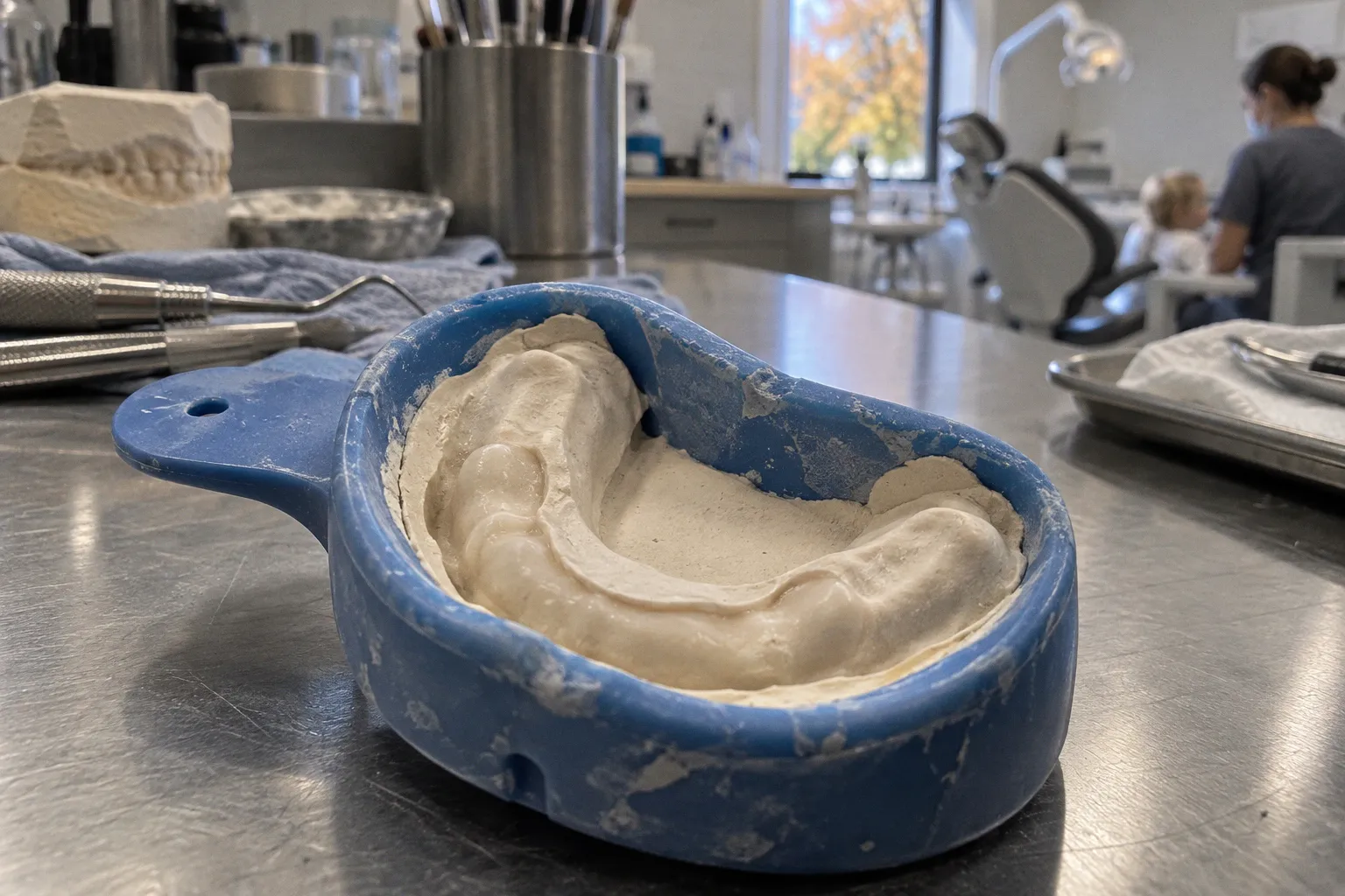
The causes behind these deviations range from genetic inheritance to systemic health conditions, and distinguishing normal variation from clinical concern requires a structured, factor-by-factor assessment.
The Biology of Eruption: Standard Timelines vs. Individual Variation
A complete primary dentition contains 20 teeth. These emerge in a predictable sequence: lower central incisors first, followed by upper central incisors, lateral incisors, first molars, canines, and second molars. The full set typically completes between 30 months and 3 years of age.
Clinical reference points for monitoring eruption:
- 6 to 12 months — typical emergence window for the first primary tooth.
- 30 months to 3 years — expected timeframe for the full set of 20 primary teeth.
- Approximately 1% of children erupt their first tooth after 12 months of age.
These figures establish a baseline, not a deadline. Practitioners assess delayed eruption against multiple variables rather than a single cutoff. A healthy infant who erupts the first tooth at 13 or 15 months may simply reflect the upper edge of normal biological variation. The clinical task is to identify which delays signal underlying pathology and which represent statistical normality within a broader population distribution.
Variation in eruption timing is the rule, not the exception — assessment must always account for the individual child's developmental context.
The Role of Genetics and Gestational Age
Genetics and family history rank among the most common non-pathological reasons for variation in primary tooth eruption timelines. When one or both parents experienced late eruption themselves, the probability that their child follows a similar pattern increases substantially. This familial pattern requires no intervention — only documentation within the child's dental record for future reference.
Premature birth and low birth weight introduce a different variable. These infants frequently demonstrate eruption schedules that align more closely with adjusted gestational age than with chronological age. A child born weeks before term may follow an eruption timeline offset by approximately the same interval. Practitioners track this by calculating the infant's adjusted age and comparing dental milestones against that reference point rather than the birth-date baseline.
This approach prevents misinterpretation of normal variation as pathology. It also allows the clinical team to monitor whether the premature infant catches up to the standard timeline over the early years, which most do without intervention. The practitioner documents gestational age, birth weight, and any neonatal complications at the first dental visit, then reassesses eruption progress at each subsequent appointment.
Systemic Health and Nutritional Drivers
Beyond genetics and gestational factors, systemic health conditions and nutritional status directly affect primary tooth development. These drivers operate at the metabolic level and produce delays that often extend beyond the oral cavity.
Endocrine disorders — including hypothyroidism and growth hormone deficiency — can disrupt the eruption sequence. These conditions affect skeletal maturation broadly, and dental development follows the same trajectory. When an infant shows delayed eruption alongside other indicators such as poor growth velocity, persistent lethargy, feeding difficulties, or constipation, endocrine screening becomes part of the clinical workup.
Nutritional deficiencies, particularly Vitamin D-resistant rickets, interfere with the calcification process required for a tooth to emerge through the gums. Vitamin D plays a central role in mineral metabolism, and insufficient availability delays the maturation that precedes eruption. Targeted supplementation, under pediatric supervision, addresses the underlying deficiency and supports subsequent dental development.
Practitioners monitor these factors through routine well-child visits, integrating dental milestone tracking with broader developmental assessment. When a child presents with delayed eruption and a documented history of the above conditions, the clinical focus expands beyond the oral cavity into coordinated care with the child's primary physician or relevant specialist.
Identifying Local Physical Obstacles
Not all eruption delays originate from systemic or genetic causes. Localized physical barriers in the oral cavity can prevent a tooth from breaking through the gums on time, even when systemic development proceeds normally.
Common local obstacles include:
- Dense fibrous gum tissue that resists the mechanical pressure of an emerging tooth and slows gingival breakthrough.
- Eruption cysts, which are fluid-filled sacs that form over an unerupted tooth and create a physical barrier at the surface.
- Arch crowding, where insufficient space in the dental arch blocks proper alignment and emergence.
- Supernumerary teeth or odontomas that occupy the eruption path and prevent normal tooth progression.
Assessment of these factors requires direct clinical examination. Practitioners palpate the gum tissue to detect hardness or swelling, assess arch dimensions through visual inspection and measurement, and — when indicated — order radiographs to visualize the position of unerupted teeth relative to the gum surface. This structured approach distinguishes a local mechanical problem from a broader developmental pattern and informs the appropriate intervention pathway.
| Factor Category | Mechanism | Typical Indicator | Clinical Response |
|---|---|---|---|
| Genetic variation | Familial inheritance pattern | Late eruption in one or both parents | Document and monitor |
| Premature birth | Adjusted gestational age offset | Low birth weight, NICU history | Track against adjusted age |
| Systemic condition | Endocrine or metabolic disruption | Reduced growth velocity, additional developmental signs | Pediatric medical workup |
| Nutritional deficit | Insufficient mineral availability for calcification | Documented Vitamin D deficiency | Targeted supplementation |
| Local obstruction | Physical barrier blocking emergence | Palpable hardness, no gingival breach | Radiographic assessment |
The 18-Month Threshold: When to Seek Professional Evaluation
Clinical guidelines recognize 18 months as the threshold for pediatric dental evaluation in an infant with no erupted teeth. This cutoff is not arbitrary — it represents the point at which the probability of an underlying developmental or structural issue rises enough to warrant formal assessment, while still allowing sufficient time for intervention if pathology is identified.
The evaluation process follows a structured protocol:
1. Medical and family history review — documenting gestation length, birth weight, parental eruption patterns, and any known systemic conditions.
2. Clinical oral examination — palpating alveolar ridges, identifying gingival abnormalities, eruption cysts, or signs of obstruction.
3. Growth and developmental assessment — comparing the child's overall developmental milestones against age-adjusted expectations.
4. Radiographic imaging — when indicated, panoramic or periapical films visualize unerupted tooth position and root development.
5. Referral pathway — if systemic conditions are suspected, the pediatric dentist coordinates with the child's primary care physician or relevant specialist.
Parents observing eruption delays before the 18-month mark should maintain a record of what they observe — dates of any gingival swelling, drooling patterns, or behavioral changes — and bring this documentation to the evaluation. Structured observation improves diagnostic accuracy and shortens the assessment timeline.
Practical Monitoring Framework
For long-term tracking of primary tooth development, practitioners and parents can follow a structured observation sequence:
- Maintain a dental milestone log from 6 months onward, noting any signs of gum activity or visible tooth tips, with dates.
- Calculate and record the infant's adjusted age if the birth was premature, then reference eruption expectations against that figure.
- Schedule the first pediatric dental visit by 12 months of age, regardless of whether teeth have emerged, to establish a clinical baseline.
- Document family eruption history — when parents and older siblings cut their first teeth provides useful context for the child's expected pattern.
- Monitor systemic health indicators — growth velocity, nutritional status, and any diagnosed endocrine or metabolic conditions.
- Seek evaluation promptly if no teeth have emerged by 18 months, or earlier if other developmental concerns coexist.
Variability in eruption timing reflects the same biological complexity that governs every other aspect of child development. Genetics, gestational age, systemic health, nutritional status, and local oral anatomy each contribute to the timeline. Practitioners assess these factors in combination, not in isolation, to distinguish normal variation from clinical concern — and to intervene when intervention produces measurable benefit for the child's long-term oral health.