Dental Innovations
3D printed dental models: visualizing your treatment plan
A 3D printed dental model is not a perfect copy of a patient’s teeth. Published measurements put the mean dimensional deviation of models made with Material Jetting at 67.8 µm and those made with stereolithography at 86.7 µm in the studies cited here.
Those numbers describe particular test conditions, not a universal guarantee for every printer, resin, or workflow. The point is not that deviation disappears. It is that it can be measured—and interpreted in context.
The move from poured stone casts to printed polymers changes how a model is made, not the need to account for tolerance. A printed model begins with a digital surface map, typically captured by an intraoral scanner, refined in CAD software, and exported for printing. The printer builds a physical object layer by layer. Each step can affect the result, from the scan and file preparation to orientation, resin, washing, and post-curing.
The Science of Precision: Understanding Dimensional Accuracy in Dental Printing
Two technologies commonly used to make dental models are Material Jetting and vat photopolymerization. In Material Jetting, print heads deposit droplets of photopolymer, which are then cured with light. Stereolithography (SLA) uses a laser to trace each layer in a vat of liquid resin. Digital Light Processing (DLP) is a related vat-photopolymerization method, but it cures a layer by projecting an image through a digital mask rather than tracing it with a laser. SLA and DLP are distinct technologies, even though both cure liquid resin layer by layer.
The resin formulations used across these processes are not necessarily the same. A shared reliance on light-activated polymerization does not establish that the materials have identical chemistry or behave identically after printing. Resin formulation, printer settings, and post-processing all matter.
In comparative measurements cited here, mean trueness error—the average geometric deviation from the digital reference—was 67.8 µm for Material Jetting and 86.7 µm for SLA. These are study results, not a ranking that holds in every clinical setup. Trueness and precision also describe different things. Trueness is closeness to the reference; precision is how consistently repeated prints reproduce the same result. A printer can be consistent without being close to the reference, or close on average while producing more variable results.
| Parameter | Material Jetting | SLA |
|---|---|---|
| Build principle | Photopolymer droplets deposited and cured | Laser traces successive layers in a resin vat |
| Reported mean trueness error in cited comparative data | 67.8 µm | 86.7 µm |
| Main sources of variation | Print settings, material, supports, and post-processing | Resin, orientation, exposure, supports, and post-processing |
A review of full-arch printed dental models reports a broad range of dimensional accuracy across SLA and DLP configurations—from under 100 µm to over 500 µm. That spread is a reminder that a technology label alone cannot predict how faithful a particular model will be. Printer class, resin, build orientation, and post-curing protocol all influence the outcome.
Light Source, Photoinitiator, and Build Orientation
Light wavelength matters because photoinitiators in the resin respond to particular wavelengths. Dental printers and materials are designed as systems: a resin’s behavior depends on the exposure conditions for which it was formulated. A clinic should therefore follow the manufacturer’s validated material and post-processing instructions rather than assume that a resin will behave the same way on any printer.
Build orientation adds another source of variation. A model printed upright may not distort in the same way as one angled on the platform. Layering, support placement, and the distribution of material across the build can affect different axes and surfaces differently. Orientation is a workflow choice, not an inherent measure of printer quality.
A model’s accuracy belongs to the whole workflow: scan, file, printer, material, orientation, and post-processing.
Clinical Standards: How 3D Models Meet ADA Restorative Requirements
A tolerance used to assess a restoration is not automatically a tolerance for a printed model. The American Dental Association’s 120 µm figure for restorative margin gaps concerns the fit between a restoration margin and a prepared tooth surface. It does not establish a pass-or-fail standard for the dimensional accuracy of a study model.
That distinction matters when interpreting tables of model measurements. A printed model with a mean deviation below a restorative margin-gap figure cannot be called compliant for crown verification, implant surgery, or another clinical use on that basis alone. The measurements concern different objects and different interfaces. Model trueness may be relevant to planning, but it cannot substitute for validating the complete procedure or verifying a restoration in the patient’s mouth.
| Question | What the measurement can tell you | What it cannot establish on its own |
|---|---|---|
| How closely does a print match its digital reference? | The model’s measured trueness under the study or workflow conditions | Whether the model is suitable for every clinical application |
| How consistently does a printer reproduce a model? | Repeatability across print cycles | Whether each print is sufficiently true to the patient’s anatomy |
| How well does a restoration fit? | Fit at the restoration–tooth interface, assessed using appropriate methods | The accuracy of a separate printed study model |
A printed study model is primarily a representation of external anatomy. If a clinician intends to use a printed model as part of restorative verification, additional checks are needed. The digital file, the printed model, and the restoration interface each have their own sources of error. A single mean trueness figure cannot collapse them into one answer.
Where Additive Manufacturing Changes the Error Profile
Stone casts and printed models do not fail in the same way. Dental stone undergoes dimensional change as it sets, with the result affected by mixing and handling. Printed polymers avoid that setting process, but they bring their own variables: layer-related artifacts, support marks, and dimensional change during curing.
The useful comparison is not that one method has no error and the other does. It is that each method has a different error profile. A model can be helpful for visualizing an arch while still being the wrong tool for a precise fit judgment. The intended use should determine how much confidence to place in its geometry.
Structural Design and Trueness: Why Cross-Arch Reinforcement Matters
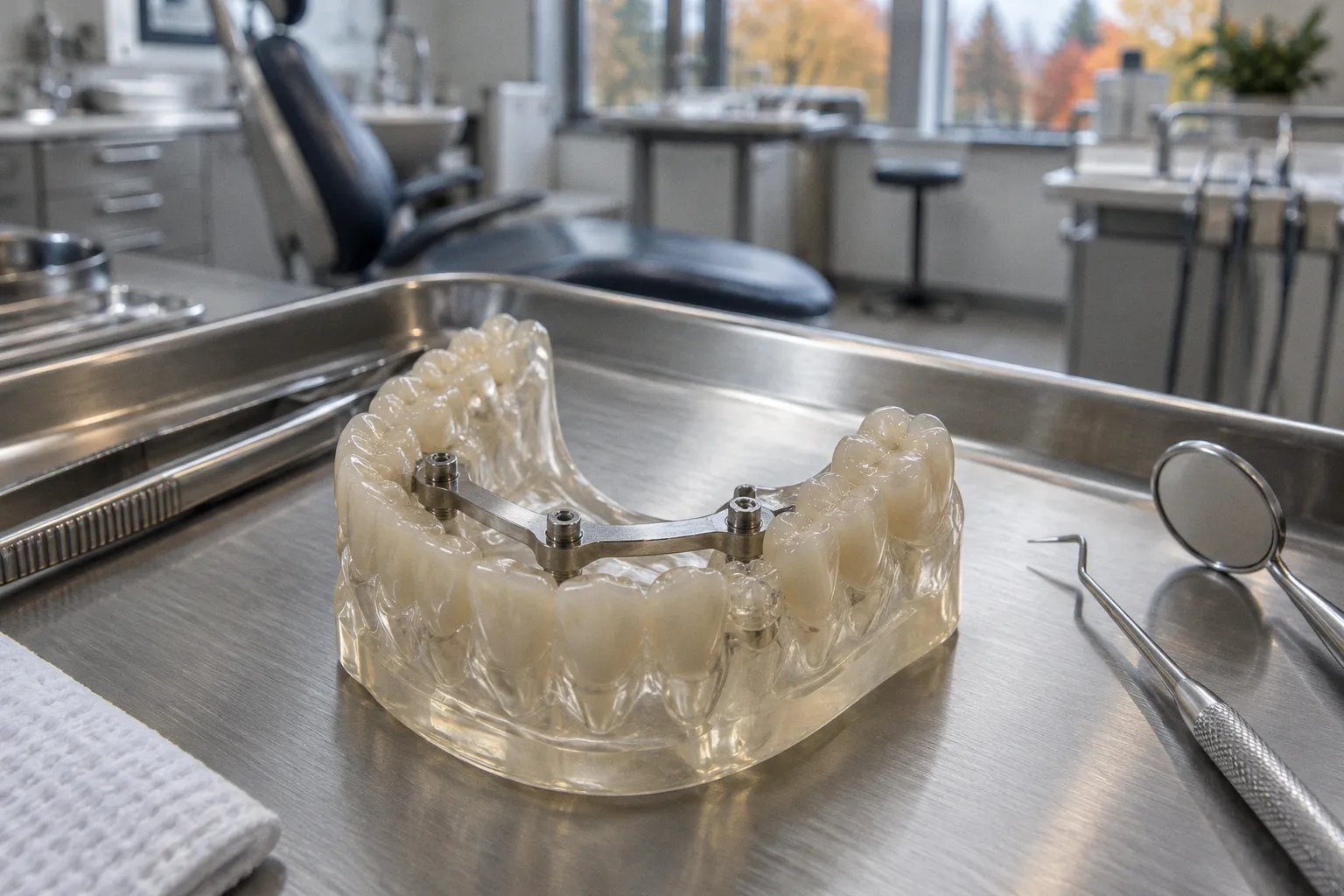
The shape of a model affects how it behaves during printing and post-curing. A U-shaped arch has an open span, and that geometry can deform more readily than a configuration reinforced across the arch. In one set of published measurements, unreinforced U-shaped models had mean trueness of 135.2 ± 26.3 µm. Models with a cross-arch reinforcement plate measured 85.6 ± 13.1 µm.
The plate connects the sides of the arch, helping resist curl and warp. In those reported results, reinforcement was associated with both a lower mean deviation and a narrower spread. That is useful evidence about the tested configuration. It is not a guarantee that adding a plate will produce the same improvement with every printer, resin, or model design.
| Model geometry | Reported mean trueness | Reported standard deviation |
|---|---|---|
| U-shaped, without reinforcement | 135.2 µm | ±26.3 µm |
| With cross-arch reinforcement plate | 85.6 µm | ±13.1 µm |
The practical point is easy to overlook: the digital model’s design is part of the manufacturing workflow. Support placement and reinforcement are not cosmetic details added after the fact. They can affect whether the printed arch keeps its shape. Where the CAD software provides reinforcement options, the choice should be made deliberately and in line with the intended use of the model.
Geometry, Supports, and Anatomical Detail
The surfaces of a dental model do not all print under the same conditions. Posterior occlusal anatomy contains pits, fissures, and concavities; these details are harder to support and reproduce consistently than flatter surfaces. Supports may leave marks, and exposure or curing may vary across complex features. Buccal and lingual surfaces can be more straightforward geometrically, but that does not make them error-free.
This is one reason a single average trueness value can be misleading. It compresses a three-dimensional pattern of deviation into one number. A model may be close to the reference overall while still showing meaningful local differences in a detail that matters for the planned conversation or procedure.
Visualizing Your Smile: Enhancing Patient Consultations with Physical Models
A digital scan gives the clinician and patient a manipulable view on a screen, but the view is mediated by the display and the selected angle. A physical model adds a different kind of access: the arch can be held, turned, and examined from multiple directions. For a conversation about orthodontic movement, a proposed implant position, or a larger restorative plan, that can make the anatomy easier to point to and discuss.
The model works best as a shared reference. A clinician can indicate where movement is planned, show how neighboring teeth relate to a proposed restoration, or explain which parts of the arch are involved. The object is scaled to the patient’s anatomy, but it remains a printed representation derived from a scan. It does not reveal structures that the scan did not capture, and it cannot answer questions that require examination or imaging beyond the tooth surfaces.
That boundary is worth stating plainly. A physical model does not replace clinical probing, radiographs, or CBCT when those are needed. It is not, by itself, a working die or proof that a final restoration will seat correctly. Its value is in making the visible geometry available for discussion—not in turning a consultation aid into a diagnostic instrument.
Research directly quantifying how much physical models change patient understanding or treatment acceptance remains limited. A tangible object may support spatial explanation, but the presence of a model alone cannot demonstrate comprehension or predict a treatment outcome. The clinician still has to connect the object to the patient’s actual options, risks, and goals.
A printed model can make a treatment plan easier to point to. It cannot make the treatment decision for the patient.
Navigating Limitations: Factors Influencing Model Fidelity and Resin Stability
The final print reflects a chain of decisions and materials. A scan can miss or soften surface detail. CAD edits can change the geometry. Printer settings, orientation, resin formulation, support placement, and post-curing can all influence the object that reaches the consultation room. When a model is used for explanation, these variables may not prevent it from being useful; they do matter when someone treats its surface as a precise measurement.
Resin behavior also depends on the formulation and its intended use. Standard model resins are used for diagnostic and presentation models. Castable resins are designed for pattern production rather than long-term retention as study models. Filled formulations may behave differently from unfilled ones, including in stiffness and processing requirements. These categories should not be treated as interchangeable.
Post-processing is part of the manufacturing process, not an optional finishing touch. Washing and curing conditions can affect the finished object’s dimensions and surface. The same digital file and printer may produce different results if processing conditions change. Evidence on long-term dimensional stability across varying humidity conditions remains limited, so long-term behavior should not be assumed from a short-term accuracy measurement.
For a patient consultation, the most defensible role for a printed model is therefore also the most useful one: a physical aid for seeing and discussing anatomy. It can help make a treatment plan concrete without being mistaken for a metrological instrument. The measurements describe how particular workflows performed; they do not grant every model a blanket approval for every clinical task.
The model’s deviation does not vanish when the file becomes an object. It changes form along the way—from scan capture to print and post-cure. Knowing that makes the model easier to use well: as a carefully bounded aid to explanation, with clinical decisions and fit verification resting on the appropriate evidence and examination.